|
|
| What are persistent pupillary membranes (PPM)?
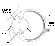
Persistent pupillary membranes are strands of tissue in the eye. They are remnants of blood vessels which supplied nutrients to the developing lens of the eye before birth. Normally these strands are gone by 4 or 5 weeks of age.
Depending upon the location and extent of these strands, they may interfere with vision. They may bridge from iris to iris across the pupil, iris to cornea (may cause corneal opacities), or iris to lens (may cause cataracts), or they may form sheets of tissue in the anterior chamber of the eye. In many dogs these tissue remnants cause no problems.
How are persistent pupillary membranes inherited?
Inheritance is not defined.
What breeds are affected by persistent pupillary membranes?
 PPM are known or strongly suspected to be inherited in the basenji, Pembroke and Cardigan Welsh corgi, mastiff, and chow chow. This problem is particularly significant in the basenji where the strands often bridge to the cornea, causing opacities which may impair sight. In the basenji the condition has been seen with optic nerve coloboma - a cavity in the optic nerve which, if large, causes blindness. PPM are known or strongly suspected to be inherited in the basenji, Pembroke and Cardigan Welsh corgi, mastiff, and chow chow. This problem is particularly significant in the basenji where the strands often bridge to the cornea, causing opacities which may impair sight. In the basenji the condition has been seen with optic nerve coloboma - a cavity in the optic nerve which, if large, causes blindness.
PPM are also seen in many other breeds, including the Akita, Alaskan malamute, American and English cocker spaniel, Australian shepherd, basset Griffin vendeen (petite), beagle, bearded collie, Belgian sheepdog, Belgian tervuren, Bichon frise, Bouviers des Flandres, Chesapeake Bay retriever, collie (rough and smooth), Doberman pinscher, English springer spaniel, golden retriever, Gordon setter, Havenese, Irish setter, Labrador retriever, Lakeland terrier, Lowchen, miniature bull terrier, Norwegian elkhound, Nova Scotia duck tolling retriever, Old English sheepdog, papillon, poodle (all sizes), Portuguese water dog, samoyed, Scottish terrier, Shetland sheepdog, soft-coated wheaten terrier, Tibetan terrier, Welsh springer spaniel, West Highland white terrier, Yorkshire terrier.
For many breeds and many disorders, the studies to determine the mode of inheritance or the frequency in the breed have not been carried out, or are inconclusive. We have listed breeds for which there is a consensus among those investigating in this field and among veterinary practitioners, that the condition is significant in this breed.
|
|
What do persistent pupillary membranes mean to your dog & you?
Generally persistent pupillary membranes cause no problems. However if attached to the cornea or lens, the strands can cause opacities which may interfere with vision. The cataracts that can occur with PPM usually don't worsen.
How are persistent pupillary membranes diagnosed?
PPM are seen in young dogs. You or your veterinarian may notice small white spots in your dog's eyes, or you may suspect that your dog's vision is impaired if the condition is severe. With an ophthalmoscope, your veterinarian will be able to see the membranous strands, and whether they adhere to the lens or cornea.
How are persistent pupillary membranes treated?
There is no treatment for the membranes themselves and in most cases there are no associated problems. If there is significant edema or "bluing" of the cornea due to adhesions, hyperosmotic eyedrops may help. Surgery may be required if there are extensive cataracts.
Breeding advice
PPM's may or may not be a problem in a breed and/or individual dogs. PPMs are remnants of a fetal structure called the pupillary membrane. This membrane covers the pupil before an animal is born. It is part of the blood supply to the developing lens (the structure in the eye that focuses light on the retina). Normally the pupillary membrane completely absorbs before birth in foals and calves but is partially present and continues to disappear in neonatal dogs. Absorption may not be complete in puppies when the eyes first open and small strands or a web-like structure may be seen across the pupil. These strands normally disappear by four to five weeks of age. In some dogs these strands do not disappear and become PPMs.
PPMs may be found in several configurations in the anterior chamber. They may span across the pupil (iris to iris), from the iris to the lens, from the iris to the cornea, or they may float free on one end, only connected to the iris. In general, iris to iris PPMs cause no problems. They may be single strands or a forked structure. These PPMs may break and become less prominent as the puppy gets older, but they usually do not disappear completely. Iris to lens PPMs are more problematical. These PPMs cause opacities (cataracts) at the point where they are attached to the lens capsule. The cataracts do not usually progress and cause only minor visual deficits. Iris to cornea PPMs cause opacities on the cornea due to their ability to damage the corneal endothelium (the inner lining of the cornea). These opacities may be small or may be severe due to the development of corneal edema (fluid in the cornea). Severely affected puppies (with numerous strands) may be blind (they may improve as they get older). The strands may regress but do not disappear.
PPMs are found in many breeds of dog. In some breeds, PPMs are known to be hereditary. The Basenji is the most well known but it is also found frequently in Chow Chows, Mastiffs, Pembroke Welsh Corgis, or Yorkshire Terriers. Members of these breeds have been shown to produce offspring with blindness directly associated with their PPMs. In these breeds, the mechanism of inheritance is not known but breeding any of these dogs with PPMs is highly discouraged.

|
RECOGNISED CERF VETERINARY OPHTHALMOLOGISTS |
|
|
| New South
Wales |
Victoria |
Dr. Cameran Whittaker
Dr. Jeff Smith
Animal Eye Clinic
104 Spofforth St.
Cremorne, N.S.W. 2090 |
Dr. Rowan Blogg
1A Irymple Av.
East Malvern,
Victoria 3146 Australia
Phone: (03) 9509 7611
|
|
|
| Queensland |
Western
Australia |
Dr. Douglas
Slatter
Dr. Elizabeth Chambers
109 Haig Road
Toowong,
Queensland 4066
Phone: (07) 3371 5763
Web: www.animaleyeclinic.com
|
Dr. Anita Dutton
Diplomate ACO
Animal Eye Clinic
855 Canning Highway
Applecross
Western Australia 6153
Phone: (08) 9315 5525 |
|
OTHER AUSTRALIAN & NEW ZEALAND EYE CLINICS |
| New
South Wales
|
New
Zealand |
Dr. Bruce Robertson,
Eyevet Associates,
274 Pennant Hills Road,
Carlingford, N.S.W. 2118
Phone: 9872-9877
|
Auckland Animal Eye Centre
18 Barrack Rd,
Mt Wellington, Auckland
Phone: 0-9-527 7697
eyevet@xtra.co.nz
|
|
|
| South
Australia |
Victoria
|
Adelaide Veterinary Specialist
and Referral Centre
Dr. R. A .Read,
102 Magill Rd, Norwood, SA 5067
Ph: (08) 8132 0533
Web: www.vetreferrals.com.au/ |
Dr Robin Stanley,
Dr Anu O'Reilly,
Dr Chloe Hardman
181 Darling Road
East Malvern Vic 3145
Phone: (03) 9563 6488
Web: www.animaleyecare.com.au
|
|
|
| Queensland |
|
Animal Eye Services
Dr Richard I E Smith, Dr Michael E Bernays, Dr Edith C G M Hampson
Cnr. Kessels Rd & Springfield St.,
Macgregor Queensland. 4109
Ph (07) 3422 2010
|
|